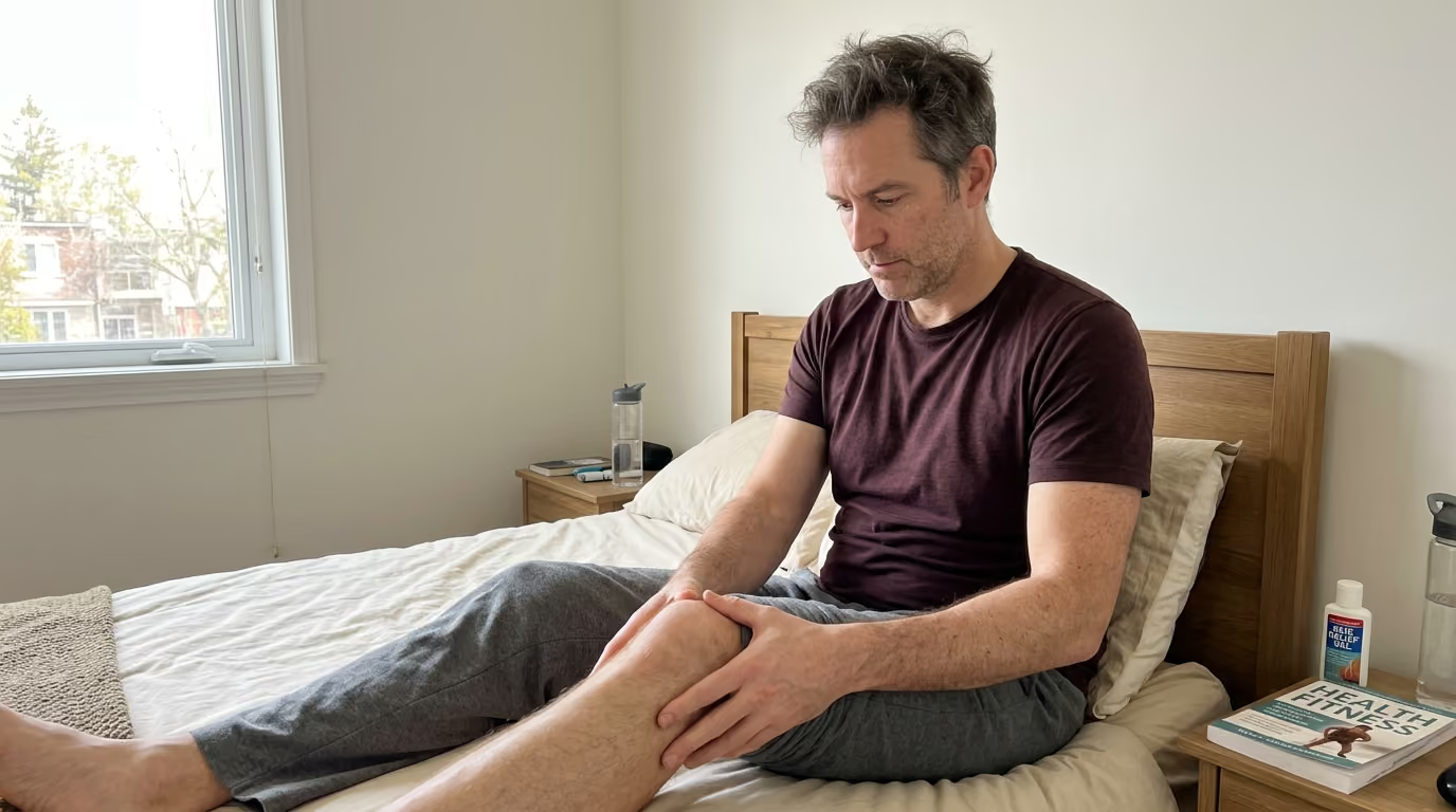

Persistent stiffness in your joints after routine exercise can be overcome through progressive load management and targeted rehabilitation protocols.





You step off the trail after a brisk morning hike in the foothills, feeling energized until a familiar ache flares in your Achilles tendon. Or perhaps it is a persistent stiffness at the base of your knee after a weekend of skiing, or a sharp twinge in your shoulder during an overhead press.
For active adults past forty, these nagging tendon issues often seem to arrive without warning and linger for months. The standard advice of taking complete rest, applying ice, and waiting for the tissue to heal on its own almost always fails.
Tendon pain is rarely an acute inflammatory event that disappears with stillness. True recovery requires understanding tendon load capacity, respecting biological remodeling timelines, and applying progressive mechanical stress to rebuild resilient tissue.
Tendon rehabilitation is a load management and capacity problem rather than an inflammation problem. When you experience tendon pain, it indicates that the mechanical demands placed on the tissue have exceeded its current load tolerance. The objective of rehabilitation is not to protect the tendon from all future stress. The real goal is to progressively restore its capacity to handle the forces of your sport, travel, and daily life.
Age serves as a risk modifier rather than an unavoidable barrier to recovery. Research indicates that mature tendons exhibit cellular changes, including fewer tendon progenitor cells and altered collagen structure. These biological shifts mean that sudden spikes in activity volume, intensity, or speed are tolerated less easily than in your twenties. However, mature tendon tissue remains remarkably responsive to mechanical loading when applied systematically.
A critical distinction must be made between persistent tendinopathy and acute tissue disruption. Tendinopathy represents a state of reduced load tolerance and structural disorganization without substantial acute inflammation. In contrast, an acute tendon tear or rupture involves mechanical discontinuity that requires medical protection or surgical repair.
Symptom relief, strength restoration, and biological tissue remodeling occur on entirely different timelines. Pain often improves within several weeks of proper load management, but structural remodeling of the collagen matrix can take a year or longer. Long-term success requires maintaining a consistent strengthening routine well after daily pain has subsided. You can explore structured conditioning approaches in our guide to strength and physical performance to maintain tissue resilience over time.
Tendons connect muscle to bone, acting as biological springs that transmit muscular force and store elastic energy during movement. Tendon tissue is predominantly composed of dense type I collagen fibers arranged parallel to the line of pull. Unlike skeletal muscle, tendons have a relatively low blood supply and a slow metabolic rate, which explains their prolonged recovery timelines.
Biological aging introduces subtle structural and cellular shifts within tendon architecture. Research published in biomechanics and sports medicine literature highlights that older tendons contain fewer functional tendon stem cells and show higher rates of collagen fragmentation. There is also an increase in non-collagenous ground substance, such as glycosaminoglycans, which can alter normal fluid dynamics and tissue stiffness. These cellular shifts reduce the baseline rate of extracellular matrix turnover.
These biological changes do not mean your tendons are degenerating irreversibly. Instead, they indicate that your tissue requires clearer signals and longer adaptation windows to respond to mechanical stress. Tendon healing follows three overlapping phases:
Because collagen cross-linking and realignment require months of progressive tension, quick fixes inevitably fail. The human body adapts specifically to the mechanical demands placed upon it. If you remove all load, the tendon down-regulates its structural stiffness and becomes weaker. If you apply progressive, controlled tension, you stimulate collagen synthesis and restore functional strength across the entire kinetic chain.
Managing tendon rehabilitation requires replacing guesswork with a systematic pain monitoring framework. Tendon pain does not provide an immediate readout of tissue damage, but it serves as a valuable gauge of recent mechanical dosage. Clinical research supports using a simple numeric scale from 0 to 10 to evaluate symptom response during and after activity.
Mild discomfort during exercise, generally rated between 0 and 3 out of 10, is acceptable and safe provided specific conditions are met. Discomfort should remain stable during the session rather than progressively worsening with every repetition. Most importantly, symptoms must settle back to your baseline within 24 hours following the session. The morning response is your most reliable test of whether the previous day's load was appropriate.
To implement this framework, track your symptoms across three distinct time points:
If your morning stiffness lasts significantly longer than usual, or if your baseline discomfort increases the following day, the previous load exceeded your current capacity. This does not mean you have failed or ruined your progress. It simply indicates that you should temporarily reduce volume, speed, or resistance before progressing again.
Understanding the difference between energy-storage loading and compressive loading is also vital. Energy-storage loads involve rapid stretch-shortening cycles, such as running, jumping, and rapid changes of direction. Compressive loads occur when the tendon wraps firmly around a bony prominence at end-range joint angles, such as deep knee flexion or deep ankle dorsiflexion. Identifying and temporarily moderating these two loading types is the fastest way to calm an irritated tendon.
Tendon rehabilitation follows a sequential progression that gradually exposes the tissue to higher forces, slower tempos, and eventually rapid elastic demands. Progressing through these stages should be determined by functional capacity and symptom stability rather than arbitrary calendar dates.
Isometric exercises involve generating muscle force without changing the joint angle or tendon length. This loading style provides a controlled mechanical stimulus with minimal joint movement and zero compressive shear stress. Clinical reviews suggest that sustained isometric holds of 45 to 60 seconds, repeated 4 to 5 times per session, can provide a short-term pain-relieving effect while maintaining muscle activation.
Isometrics are an entry point during high-irritability phases rather than a standalone cure. They allow you to load the muscle-tendon unit when dynamic movements are too painful. As soon as symptoms stabilize, you should introduce dynamic strength work.
Heavy slow resistance forms the foundation of long-term tendon recovery. This stage uses slow concentric and eccentric phases, typically lasting 3 to 4 seconds up and 3 to 4 seconds down. The controlled tempo eliminates rapid momentum, reducing high-impact peak forces while maximizing mechanical tension across the collagen matrix.
A randomized clinical trial comparing heavy slow resistance to isolated eccentric training in Achilles tendinopathy demonstrated comparable improvements in VISA-A scores and tissue thickness at 12 and 52 weeks. Heavy slow resistance protocols often yield higher patient satisfaction because they require fewer weekly sessions while building substantial muscular strength. Perform these exercises 2 to 3 times per week, allowing at least 48 hours of recovery between sessions to support collagen synthesis.
Once heavy slow resistance is tolerated with minimal next-day symptoms, you must reintroduce the tendon to rapid elastic loading. Tendons act like springs during dynamic activity, storing and releasing kinetic energy within fractions of a second. Slow resistance training builds capacity, but dynamic movement teaches the nervous system and tendon to handle high-velocity strain.
Begin with low-amplitude elastic tasks, such as double-leg hopping or skipping drills, before progressing to single-leg bounds, deceleration drills, and directional changes. Keep the volume low initially, tracking the 24-hour response carefully before increasing speed or repetition counts.
The final phase bridges the gap between structured rehabilitation and unrestricted sport. In this stage, you reintroduce the specific volumes, velocities, and movement patterns required by your favorite pursuits. A runner must build sustained mileage, a tennis player must handle repetitive serving and stopping, and a skier must tolerate sustained isometric quad burn with sudden eccentric absorbing loads.
Readiness is determined by objective functional tests, such as symmetry in single-leg strength and hop capacity, rather than the complete absence of resting awareness. You can find comprehensive recovery frameworks in our resource on healthy aging and living well to support this transition.
Different anatomical regions impose unique biomechanical stresses on their respective tendons. Tailoring your rehabilitation to the specific demands of each tendon site ensures optimal recovery.
Achilles issues are typically categorized as midportion or insertional tendinopathy. Midportion problems occur two to six centimeters above the heel bone, where blood supply is relatively low. Insertional problems occur directly at the calcaneus attachment and are highly sensitive to compressive loading in deep dorsiflexion.
For midportion tendinopathy, begin with standing straight-knee calf raises and seated bent-knee calf raises to target both the gastrocnemius and soleus muscles. Progress from bilateral flat-ground raises to heavy single-leg heel raises off the edge of a step. For insertional tendinopathy, perform all initial heel raises from flat ground to avoid compressing the tendon against the heel bone. Gradually introduce deficit heel raises off a step only after resting symptoms and morning stiffness have resolved.
A 52-year-old runner experiencing morning Achilles stiffness should reduce running volume, replace speedwork with flat zone-2 jogging, and initiate heavy slow heel raises three times per week. Monitor the single-leg heel-rise capacity and morning stiffness duration to guide progression back to hill repeats and interval training.
Patellar tendinopathy, often called jumper's knee, manifests as localized pain at the inferior pole of the patella. It is provoked by movements involving deep knee flexion combined with rapid quadriceps deceleration, such as jumping, downhill hiking, or deep squatting.
Early management centers on reducing provocative jumping and deep impact while introducing sustained knee extensions or Spanish squats. Spanish squats use a heavy resistance band looped behind the knees, allowing a vertical shin angle that creates high quadriceps load with minimal patellar compression.
A landmark randomized trial published in the British Journal of Sports Medicine compared progressive tendon loading with eccentric exercise therapy over 24 weeks. The progressive loading group achieved a 28-point improvement on the VISA-P scale compared to an 18-point improvement in the eccentric group. Progress from isometric knee extensions to heavy slow leg presses, goblet squats, and eventually forward deceleration lunges.
Proximal hamstring tendinopathy presents as deep, localized ache at the ischial tuberosity, often referred to as the sitting bone. This condition is aggravated by combined hip flexion and hamstring tension, which compresses the tendon against the pelvis. Common triggers include deep lunges, low deadlifts, hill running, and prolonged sitting on hard chairs.
Initial rehabilitation requires limiting deep hip flexion and avoiding aggressive hamstring stretching. Begin with isometric supine bridge holds, positioning the knees at a 90-degree angle with neutral hip alignment. A standard protocol includes 5 sets of 45-second holds performed daily or every other day.
As symptoms stabilize, progress to long-lever bridges, single-leg romanian deadlifts with limited range, and slow lying hamstring curls. Only introduce deep hip flexion and high-speed sprinting after the tendon tolerates heavy resistance without next-day pain.
Rotator cuff tendinopathy involves chronic overload of the supraspinatus, infraspinatus, or subscapularis tendons. It manifests as pain during overhead reaching, lateral arm elevation, or sleeping on the affected side. A thorough clinical assessment is essential to differentiate tendinopathy from full-thickness tears, frozen shoulder, or cervical radiculopathy.
Clinical practice guidelines emphasize progressive strengthening of the rotator cuff and scapular stabilizers. Begin with pain-tolerated isometric external rotation and abduction against a wall or light resistance band. Progress to side-lying external rotations, cable face pulls, and dumbbell scaption raises within a comfortable range of motion.
Incorporate dynamic stability drills, such as kettlebell bottoms-up carries, to train shoulder endurance and neuromuscular coordination. Ensure that overhead lifting volume is managed conservatively until basic external rotation strength is symmetrical with the unaffected side.
Lateral elbow tendinopathy, commonly known as tennis elbow, affects the common extensor origin at the lateral epicondyle, primarily involving the extensor carpi radialis brevis. It is triggered by repetitive wrist extension, forceful gripping, racquet sports, or prolonged keyboard and mouse use.
Rehabilitation requires building capacity in the forearm extensor musculature while managing daily gripping loads. Begin with supported isometric wrist extensions using a dumbbell or elastic resistance bar. Hold the wrist in slight extension for 30 to 45 seconds across 4 to 5 repetitions.
Progress to slow concentric and eccentric wrist extensions, forearm pronation and supination, and isolated grip strength training. A critical error in elbow rehabilitation is neglecting the shoulder and upper back. Strengthening the rotator cuff and scapular retractors reduces the compensatory mechanical burden placed on the smaller forearm muscles during manual tasks and sports.
Overcoming tendon pain requires unlearning several pervasive myths that derail recovery and delay return to activity.
The most damaging misconception is that complete rest will heal a tendon. While resting for several weeks may temporarily reduce pain, it also decreases muscle volume, reduces tendon stiffness, and diminishes overall physical capacity. When you eventually return to activity, the tendon is even less capable of handling the load, resulting in a swift recurrence of symptoms. Active, progressive loading is required to stimulate matrix remodeling.
Passive therapies such as ultrasound, massage, acupuncture, and ice packs may offer brief pain relief, but they do not alter tendon load capacity. Relying on passive modalities without a structured strengthening plan leads to chronic frustration. Passive treatments should only serve as minor adjuncts to progressive resistance training.
Magnetic resonance imaging and ultrasound scans frequently reveal structural abnormalities, such as tendon thickening or partial collagen tears, in completely pain-free active adults. Research consistently shows that structural imaging does not correlate perfectly with pain levels or functional ability. Base your rehabilitation progress on your strength, movement quality, and next-day symptom response rather than repeated scan results.
While isometric exercises are valuable for managing acute irritability and building early confidence, they do not build complete functional capacity. Isometrics do not train dynamic stretch-shortening cycles or eccentric force absorption. Relying solely on isometric holds leaves the tendon unprepared for the demands of running, jumping, and athletic movement.
Accepting mild discomfort during exercise is necessary, but ignoring progressive flares is counterproductive. If your pain escalates with each successive set, or if your morning stiffness worsens day after day, you are overloading the tissue. Respect the 24-hour response rule and adjust your training volume accordingly.
You do not need to spend hours each day on complicated physical therapy routines to achieve robust tendon recovery. A streamlined, highly focused protocol performed consistently produces superior results while fitting into a demanding schedule.
Focus on two to three dedicated strengthening sessions per week, spaced 48 to 72 hours apart. Each session should contain two to three targeted exercises performed with high concentration and strict tempo.
On non-lifting days, engage in low-impact aerobic exercise such as cycling, swimming, or flat walking to maintain cardiovascular fitness and promote systemic circulation. Prioritizing consistent recovery and regeneration ensures that collagen synthesis and tissue remodeling occur efficiently between loading bouts.
Track your recovery using validated functional measures rather than daily pain variations. For Achilles issues, track your single-leg heel-rise repetitions and the VISA-A questionnaire monthly. For patellar problems, monitor your single-leg decline squat tolerance and the VISA-P score. Consistent improvement in these functional metrics confirms that your tendon capacity is expanding.
International travel and mountain sports introduce unique mechanical stresses that can irritate vulnerable tendons. Prolonged flights, heavy luggage handling, steep alpine ascents, and unaccustomed walking on cobblestones can rapidly spike tendon loads.
During long-haul flights, prolonged sitting with bent knees and hips increases compressive stress on the patellar and proximal hamstring tendons. Stand up and walk the aisle every two hours to relieve joint compression and stimulate circulation. Perform discreet isometric calf contractions and seated ankle rotations to maintain tissue mobility during transit.
When planning demanding active travel, prepare your tendons well in advance:
If you experience a tendon flare while traveling in remote terrain, modify your movement patterns immediately. Shorten your stride on uphill sections to reduce Achilles tension, use trekking poles on descents to offload the patellar tendons, and avoid deep stretching in the evening. Maintain mild daily movement to prevent joint stiffness rather than remaining completely sedentary in your lodge. You can find more strategies for staying resilient on the road in our travel and adventure section.
The broader sports medicine and orthopedic community agrees on several core principles regarding tendon management in mature adults. Major clinical practice guidelines from the Academy of Orthopaedic Physical Therapy and the Journal of Orthopaedic & Sports Physical Therapy emphasize progressive loading, individualized load management, and objective functional assessment over passive interventions.
While conservative loading is the gold standard for chronic tendinopathy, specific warning signs warrant an immediate medical evaluation. Consult an orthopedic specialist or sports medicine physician if you experience any of the following:
When managed with patience, scientific rigor, and consistent mechanical loading, mature tendons retain an exceptional capacity to adapt, strengthen, and support a lifetime of high-level adventure.
Revisit this guide whenever you plan to significantly increase your training volume, prepare for a strenuous travel expedition, or experience the initial onset of localized joint or tendon stiffness.
With a structured loading program and disciplined daily monitoring, you can build resilient, capable tendons that keep you moving with confidence across every decade.
Stay connected for research and practical guidance on longevity, strength, recovery, energy and active living. New ideas to help you stay capable, curious and ready for more.


Explore practical guidance on strength, recovery, energy, travel and longevity for a life that stays active.
explore the Blog