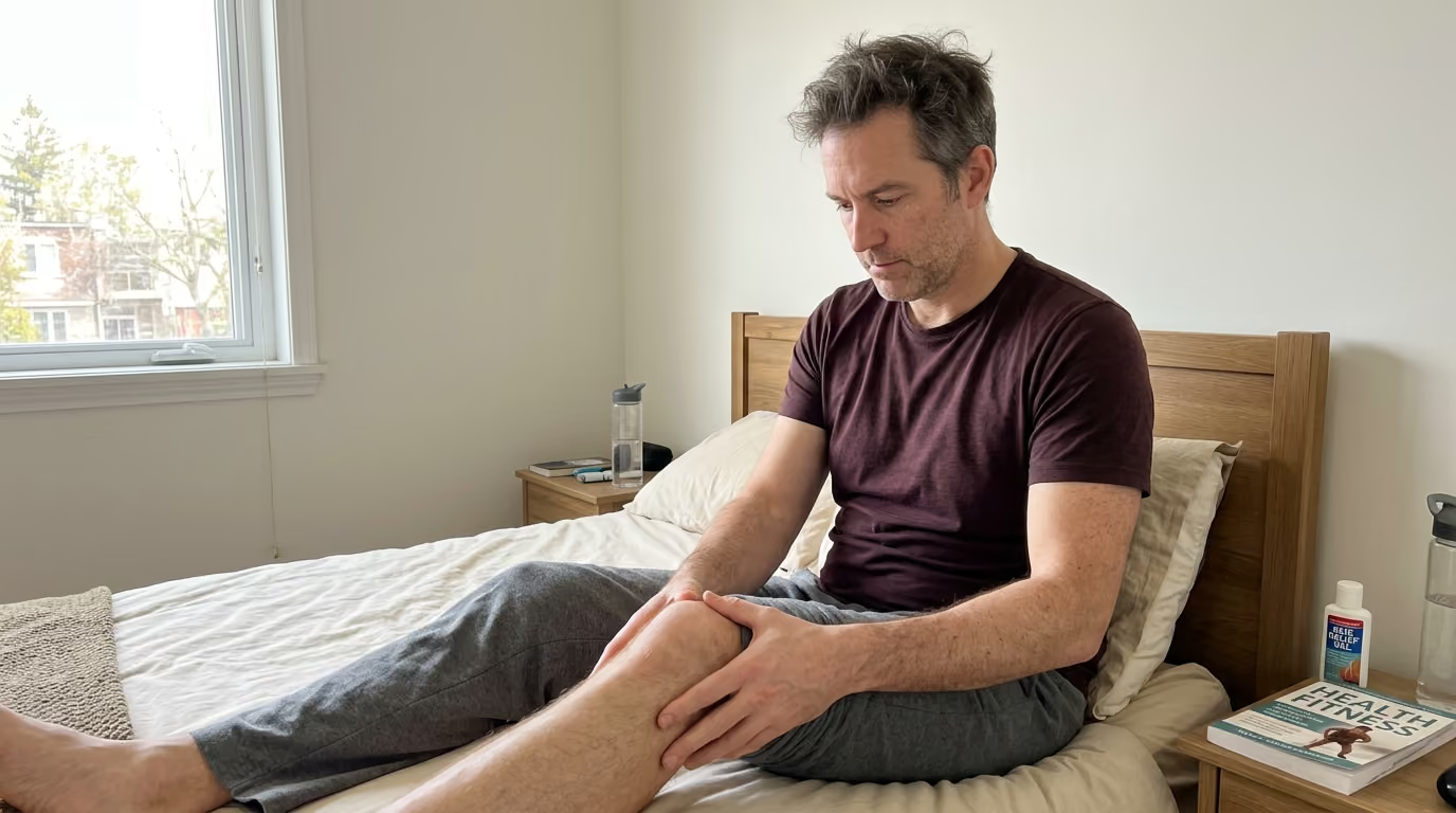

Pain intensity often masks true tissue damage, but systematic triage of timing, location, and functional capacity accurately distinguishes benign soreness.





Post-exercise discomfort is not a single diagnosis, nor is it a simple badge of athletic honor. It is a biological feedback signal that reflects how muscles, tendons, joints, and nerves respond to mechanical tension and metabolic stress. Distinguishing between normal muscular remodeling and an incipient tissue injury is not about guessing pain tolerance. It is a structured process of pattern recognition that evaluates timing, anatomical location, load response, functional capacity, and symptom trajectory.
Active adults who regularly ski, lift, run, or travel need a clear framework to interpret these physical signals. Misinterpreting normal delayed soreness as an acute injury can lead to unnecessary fear and excessive rest. Conversely, ignoring focal tendon pain, structural joint instability, or systemic distress can transform a minor, manageable issue into a chronic setback. This comprehensive guide outlines the underlying physiology of exercise-induced discomfort, provides a six-point screening protocol, and establishes clear clinical red flags to help you protect long-term physical capability.
Delayed-onset muscle soreness is a benign, self-limiting biological response to unfamiliar or demanding muscular work. It represents microstructural remodeling in muscle fibers and connective tissue. It typically emerges after a pain-free window of twelve to twenty-four hours, reaches peak intensity between twenty-four and seventy-two hours, and resolves within roughly a week. The discomfort is usually diffuse, bilaterally symmetrical when training both sides, and confined to the main body of the working muscles.
Tissue injury operates under completely different biological rules. An acute muscle strain, ligament sprain, tendinopathy, or joint pathology involves structural failure or localized tissue overload. Symptoms frequently begin during a specific movement, acceleration, or landing. They can also accumulate insidiously at a single anatomical attachment point over weeks of unmanaged volume.
The most dependable distinction is not how much an area hurts. Pain intensity is notoriously deceptive. A mild, painless loss of joint stability or subtle nerve tingling can indicate a more urgent issue than severe, widespread quadriceps soreness after a heavy session of lunges. The essential difference lies in functional preservation and symptom behavior. Normal soreness allows you to walk, hinge, and perform daily tasks once you warm up, even if you feel stiff. A true injury impairs basic mechanics, worsens under direct load, and fails to follow a predictable trajectory of daily improvement.
Understanding these recovery signals is central to structured recovery and regeneration. Developing sharp self-monitoring skills allows active adults to make informed training adjustments without losing momentum.
To interpret physical feedback accurately, you must understand the distinct physiological mechanisms behind delayed soreness, acute strains, chronic tendon irritation, joint inflammation, nerve compression, and systemic muscle breakdown.
Delayed-onset muscle soreness, commonly abbreviated as DOMS, is primarily triggered by unaccustomed eccentric contractions. Eccentric loading occurs when a muscle lengthens while producing force, such as lowering a heavy dumbbell or descending a steep mountain trail. Research published in the Journal of Physiology demonstrates that eccentric contractions place high mechanical tension on individual muscle fibers. This tension produces microscopic disruptions in the structural sarcomeres and the surrounding extracellular matrix.
This structural disruption initiates a local inflammatory cascade. Research reviewed in Frontiers in Physiology confirms that immune cells migrate to the affected muscle tissue over the subsequent twenty-four to forty-eight hours. These cells release bradykinin, prostaglandins, and histamine, which sensitize local type III and IV sensory nerve endings. This biological timeline explains why you rarely feel sore immediately after finishing a session. The sensation peaks only after the inflammatory signaling reaches its biological maximum. As the muscle fibers repair and remodel, force production returns to normal, leaving the tissue more resilient to identical future workouts.
An acute muscle strain is a physical tear of muscle fibers or the musculotendinous junction, where muscle transitions into tendon. Unlike the microscopic, diffuse microtrauma of DOMS, a strain involves macroscopic structural damage caused by acute mechanical failure. Strains frequently happen when a muscle is forced to contract while being rapidly stretched, such as during a sprint acceleration or an awkward slip on uneven terrain.
The resulting trauma ruptures local capillaries, leading to internal bleeding, localized swelling, and inflammatory fluid accumulation. The nervous system responds immediately with protective muscle spasm to splint the injured region. Pain from an acute strain is almost always sharp, sudden, and strictly localized to the injured site.
Tendons connect muscle to bone, transmitting massive tensile forces during movement. Tendinopathy is a broad clinical term for tendon pain and impaired load tolerance. Unlike acute tendon ruptures, most tendinopathies develop gradually due to repetitive loading that exceeds the tendon cellular repair capacity.
Healthy tendons consist of tightly packed, parallel collagen fibers. When subjected to excessive volume, rapid spikes in training frequency, or poor movement mechanics, tendon cells enter a reactive state. As detailed in the British Journal of Sports Medicine, reactive tendinopathy involves localized swelling, matrix disruption, and increased sensitivity of local nociceptors.
Tendons possess unique mechanical behavior known as the warm-up effect. A reactive Achilles or patellar tendon often feels stiff and painful during the first ten minutes of morning walking or early in a workout. As tissue temperature and blood flow increase, the pain temporarily subsides, creating a false impression of full recovery. However, once the session ends and the tendon cools, symptoms return with greater intensity later that evening or the following morning.
Joint discomfort originates within the articular cartilage, synovial membrane, joint capsule, or surrounding stabilizing ligaments. Cartilage itself is aneural, meaning it lacks sensory nerve fibers. Pain associated with joint irritation arises when mechanical wear, poor alignment, or sudden impact stresses the sensitive synovial lining, subchondral bone, or joint capsule.
When a joint is irritated, the body produces excess synovial fluid, causing intra-articular swelling. This swelling reduces joint range of motion and reflexively inhibits the surrounding musculature. For example, knee effusion quickly shuts down full activation of the vastus medialis oblique muscle. Joint pain is typically experienced deep within the joint space, along the joint margin, or as a sensation of mechanical catching, clicking, or instability.
Post-exercise discomfort does not always stem from musculoskeletal tissue. Peripheral nerves pass through narrow anatomical tunnels formed by muscle, fascia, and bone. Excessive muscular tightness, joint inflammation, or spinal disc irritation can compress or stretch these neural pathways.
Nerve symptoms possess distinct qualitative markers. Rather than a dull ache or local tenderness, neural irritation produces burning sensations, electric shocks, numbness, or pins and needles. These symptoms often radiate distal to the site of compression, traveling down an arm or leg. Nerve compression can also cause genuine neurological weakness, where a limb feels disconnected or fails to fire regardless of voluntary effort.
Exertional rhabdomyolysis is a serious, potentially life-threatening medical condition caused by massive, rapid breakdown of damaged skeletal muscle tissue. While DOMS involves minor structural remodeling, rhabdomyolysis represents widespread myocyte cell death. When muscle cell membranes rupture, large volumes of intracellular contents, including potassium, electrolytes, and the protein myoglobin, spill into the bloodstream.
Myoglobin is filtered through the kidneys, where high concentrations can cause acute tubular necrosis and kidney failure. Clinical practice guidelines published in the American College of Sports Medicine literature note that exertional rhabdomyolysis is characterized by serum creatine kinase (CK) levels rising at least five times above the upper limit of normal, often exceeding 5,000 to 30,000 IU/L. This condition is triggered by extreme, unaccustomed high-volume exercise, especially in hot conditions or when compounded by dehydration.
Self-monitoring should never rely solely on a one-to-ten pain rating. Pain intensity fluctuates based on stress, sleep, and personal perception. A far more accurate approach is a systematic six-question screen that evaluates symptom behavior, mechanical capacity, and systemic health.
The exact moment symptoms appear is a powerful clinical clue. Delayed-onset muscle soreness follows a predictable physiological arc. You finish your training feeling fatigued but relatively comfortable. Noticeable stiffness and tenderness begin twelve to twenty-four hours later, peaking between forty-eight and seventy-two hours before gradually declining.
If discomfort begins during a specific repetition, sprint, or landing, you are likely dealing with an acute strain, sprain, or joint subluxation. Immediate pain suggests acute mechanical disruption of structural fibers. Alternatively, if discomfort starts insidiously across several weeks, appearing midway through a run or climb, it points toward a chronic overuse issue like tendinopathy or a bone stress injury.
Anatomical location provides essential diagnostic context. Delayed soreness is broadly distributed across the muscle bellies that performed mechanical work. If you performed back squats, both quadriceps and gluteal muscles will feel uniformly tender across their broad muscular surfaces.
Injury patterns are far more focal and asymmetric. Ask yourself if you can touch the exact epicenter of the pain with the tip of a single finger. Pinpoint tenderness directly over a bony prominence, a tendon insertion, or along a joint line suggests localized structural pathology. Pain that radiates down a limb or follows a specific nerve path indicates spinal or peripheral nerve involvement.
The qualitative character of the pain reflects the underlying tissue type. Benign muscle soreness feels like a heavy, dull ache accompanied by generalized stiffness and mild tenderness when the muscle is firmly pressed. It often feels pleasant to gently move, massage, or warm the tissue.
In contrast, sharp, stabbing, or tearing sensations point toward structural tears in muscle fibers, tendons, or ligaments. Sensations of mechanical locking, catching, or giving way indicate intra-articular pathology, such as a meniscus or labral tear. Sensations of heat, burning, stinging, or pins and needles signal that nerve tissue is compressed or irritated.
How tissue responds to movement separates healthy adaptation from persistent overload. When you begin light movement with standard DOMS, the muscle initially feels stiff. However, as blood flow increases and tissue temperature rises, range of motion improves and the ache recedes.
Injured or reactive tissue behaves very differently. Tendons and joints may tolerate light, flat movement once warm, but they react negatively to higher speed, explosive stretch-shortening cycles, or deep joint angles. Clinical guidelines from the Ohio State University Sports Medicine team highlight a critical load monitoring rule: if a training session causes pain that exceeds baseline and remains elevated twenty-four hours later, that session exceeded tissue capacity. Persistent next-day aggravation is a clear sign to modify training.
Evaluating physical function is far more reliable than assessing pain intensity alone. Compare your affected side with your unaffected side during basic movement patterns. Stand on one leg, perform an unweighted bodyweight squat, walk down a flight of stairs, or reach overhead.
True muscle soreness may make these tasks feel uncomfortable, but your fundamental biomechanical capacity remains intact. You can still support your body weight, achieve standard joint angles, and control your balance. If you find yourself limping, unable to bear weight, lacking normal joint extension, or experiencing sudden muscular weakness, you are dealing with functional failure. Such limitations indicate an acute injury or joint blockage that requires rest and professional assessment.
A biological recovery signal must always be evaluated across time. Plotting the trajectory of your symptoms across several days provides clarity that a single snapshot cannot.
A healthy recovery trajectory is consistently positive. Even if DOMS is severe at forty-eight hours, symptoms are noticeably better by day four, and the muscle feels normal by day six or seven. If symptoms remain completely static for more than a week, or if they steadily intensify with each passing day despite rest, the tissue is not resolving on its own. An escalating or non-resolving trajectory demands an immediate step down in training volume and professional medical guidance.
Understanding the differences between post-exercise conditions helps prevent misdiagnosis and guides smart training adjustments.
To see these diagnostic principles in action, examine how recovery signals manifest in typical scenarios experienced by active adults over forty.
A fifty-two-year-old executive returns to the gym after a month of travel, completing a high-volume session of barbell squats and walking lunges. Two days later, both thighs feel bruised, heavy, and intensely stiff. Climbing stairs is uncomfortable, and sitting down into a chair requires a conscious effort.
This presentation matches textbook delayed-onset muscle soreness. The stimulus was unaccustomed eccentric loading. Symptoms appeared after a twenty-four-hour delay, are distributed symmetrically across both quadriceps, and involve no joint swelling or bruising. The individual can still bear weight and walk without a limp. Light cycling and walking will increase blood flow and accelerate functional recovery.
A forty-six-year-old recreational soccer player sprints toward a loose ball during a weekend match. Fifty meters into the sprint, he feels a sudden, sharp pinch in the middle of his right hamstring, forcing him to decelerate immediately. The following morning, the hamstring is tender to the touch, and walking with a normal stride causes a sharp catching sensation.
This is an acute muscle strain. The sudden onset during a high-velocity movement, the strict unilateral focus, and the functional gait change rule out standard DOMS. Attempting to stretch the muscle aggressively will only worsen the structural tear. This individual requires active relative rest, temporary load modification, and a progressive rehabilitation protocol focused on eccentric remodeling.
An avid trail runner in her late forties increases her weekly mountain elevation gain to prepare for an alpine trek. A week later, she notices that her right Achilles tendon feels extremely stiff and tender during her first twenty steps out of bed each morning. The tendon loosens up after a hot shower, allowing her to run without major distress, but throbs that evening.
This pattern is classic reactive tendinopathy. The progressive onset, localized tendon pain, morning stiffness, and the deceptively comfortable warm-up window are hallmark signs. Pushing through this phase will degrade the tendon matrix into chronic degeneration. The solution involves temporarily reducing uphill running volume, eliminating high-speed plyometric loading, and introducing heavy, slow resistance training to rebuild tendon stiffness.
Applying targeted loading protocols is an essential part of strength and physical performance as we age.
A forty-three-year-old cross-training athlete incorporates high-repetition box jumps into his routine. The following afternoon, he experiences sharp pain localized directly behind his kneecap whenever he walks down stairs or lowers into a squat. The front of his knee appears puffy, and pressing his finger along the inferior edge of the patella reproduces the sharp pain.
This scenario represents patellar tendon irritation combined with patellofemoral joint stress. The isolated joint-line pain, visible intra-articular swelling, and specific provocation during compressive knee flexion are incompatible with diffuse muscular DOMS. The athlete needs to eliminate jumping and deep loaded knee flexion, substituting flat isometric exercises while assessing hip and ankle mechanics.
A forty-eight-year-old traveler joins an intense, high-repetition kettlebell class in a humid resort environment after several months of minimal physical conditioning. Twenty-four hours later, her arms and shoulders are swollen, rock-hard to the touch, and so weak that she cannot lift a glass of water. Later that evening, she notices that her urine is the color of dark iced tea.
This is an urgent medical red flag for exertional rhabdomyolysis. The extreme muscular swelling, profound functional weakness, and dark urine point to extensive muscle cell breakdown and myoglobinuria. This situation requires immediate emergency medical evaluation, intravenous hydration, and clinical monitoring to protect renal function.
A fifty-year-old lifter attempts a series of heavy overhead barbell presses. The next morning, he feels an ache in his neck and upper shoulder, accompanied by an electric, tingling sensation traveling down his arm into his thumb and index finger. His grip strength feels noticeably weak when picking up a briefcase.
This presentation indicates cervical nerve root compression or brachial plexus irritation. The radiating quality, pins and needles sensation, and measurable grip weakness confirm neural involvement rather than simple trapezius muscle soreness. Overhead pressing should cease immediately until a physical therapist or physician evaluates the cervical spine.
A seasoned master marathoner increases his training volume while managing high workplace stress and irregular sleep. Over four weeks, his running times steadily deteriorate despite working harder. He wakes up every morning with heavy, achy legs, unrefreshing sleep, an elevated resting heart rate, and profound mental irritability.
This is not localized musculoskeletal damage, but systemic non-functional overreaching bordering on overtraining syndrome. As outlined in consensus statements from the European College of Sport Science and the American College of Sports Medicine, excessive training stress without adequate recovery causes systemic neuroendocrine and immune dysregulation. The solution requires a comprehensive deload, nutritional optimization, and dedicated sleep restoration.
Maintaining metabolic balance and physical capacity is central to healthy aging and living well.
Flawed assumptions about training discomfort cause unnecessary injuries and disrupt athletic consistency. Clearing away these misconceptions allows you to interpret your physical signals with clinical precision.
Many active adults still believe that the success of a training session is measured by how sore they feel the following day. Sports science has thoroughly disproven this idea. DOMS is simply a marker of novelty and unaccustomed eccentric work, not a mandatory driver of muscular hypertrophy or cardiovascular adaptation. Highly trained athletes frequently make exceptional progress in strength, muscle mass, and endurance while experiencing minimal post-exercise soreness. Chasing extreme soreness usually leads to excessive tissue damage, prolonged recovery timelines, and compromised training consistency.
Visual appearance can be deeply misleading. While high-grade muscle tears and severe ligament ruptures often produce visible ecchymosis, many significant injuries show no surface bruising at all. Deep muscle strains, intra-articular cartilage tears, labral damage, and early bone stress injuries rarely produce visible discoloration. Relying on the absence of bruising to clear yourself for hard training is a dangerous mistake. Functional movement capacity and load tolerance are far more dependable indicators of tissue health.
This is the classic tendon trap. Reactive tendinopathy is notorious for easing during the initial phases of low-speed movement as tissue temperature rises. Athletes often interpret this temporary relief as a green light to perform sprint work, plyometrics, or heavy lifting. In reality, the tendon is accumulating microtrauma that will trigger a severe inflammatory spike hours later. If a painful area requires ten to fifteen minutes of warming up just to feel tolerable, the underlying tissue is compromised and requires load reduction.
When muscles feel tight and restricted after training, the instinctive reaction is often to perform prolonged, aggressive static stretching. If that tightness is caused by an acute micro-tear or structural strain, pulling the muscle into extreme lengthened positions places tensile stress across damaged fibers. This can worsen structural tearing and delay healing. Tightness is frequently a protective neurological response designed to prevent further movement. Gentle active mobility and light movement are far safer and more effective than aggressive static stretching.
Modern wearable rings, watches, and bands provide useful data regarding resting heart rate, heart rate variability, and sleep architecture. However, they cannot assess the structural integrity of your peripheral tissues. A wearable device might display an optimal recovery score based on autonomic balance while your patellar tendon is in an acute reactive state. Wearable metrics should serve as secondary context, never as a replacement for physical self-assessment and symptom monitoring.
Discovering how to balance training metrics with internal physiological signals is a core focus across WealthAtPlay, where capability takes precedence over blind tracking.
Managing recovery signals does not require complicated routines or expensive equipment. It requires an actionable decision-making system that guides your daily training volume based on real-time tissue feedback.
To eliminate guesswork, categorize your daily physical recovery signals into three distinct operational tiers.
Before starting any training session, take three minutes to run through a standardized physical check. This simple routine captures early warning signs before they escalate into significant setbacks.
First, perform five slow, unweighted bodyweight squats, paying attention to bilateral symmetry, knee tracking, and hip comfort. Second, perform a single-leg balance and calf raise on each side, assessing Achilles tendon stiffness and ankle stability. Third, reach both arms fully overhead and rotate your thoracic spine in both directions to confirm clear joint mechanics and spinal comfort. If any step reveals focal pain, asymmetry, or restricted movement, adjust your workout before lifting a single weight.
Smart self-monitoring helps you maintain consistent physical progress. For broader strategies on managing lifestyle, training, and longevity, explore our comprehensive healthy aging articles.
For active adults who prioritize travel, mountain sports, and international adventure, interpreting physical signals becomes more complex. Travel introduces distinct physiological stressors that amplify discomfort and distort standard recovery timelines.
Long-haul flights and extended vehicle transit place the musculoskeletal system in prolonged, static positions. Sitting for eight to twelve hours in a cramped airplane seat restricts lower-limb circulation, promotes venous pooling, and increases passive fluid retention around the ankles and knees. The hip flexors and hamstrings remain in shortened positions, while the lumbar spine endures continuous compressive load.
Furthermore, aircraft cabin environments have low humidity levels, typically between ten and twenty percent, which accelerates subclinical dehydration. Dehydration reduces blood plasma volume, impairs nutrient delivery to healing tissues, and reduces the cushioning capacity of articular cartilage. When you land and immediately head to the hotel gym or hit the pavement for a run, your tendons and spinal discs are less compliant, significantly increasing the risk of acute irritation.
Trekking in high-altitude environments like the Alps, the Rockies, or the Himalayas presents unique physiological challenges. At altitudes above 8,000 feet, reduced ambient oxygen pressure lowers arterial oxygen saturation. This systemic hypoxia impairs cellular energy production and slows the clearance of metabolic byproducts, making ordinary physical exertion feel significantly harder and extending the duration of post-workout fatigue.
Mountain sports also place extraordinary eccentric demands on the lower body. Descending steep, rocky trails requires continuous eccentric braking from the quadriceps, patellar tendons, and calves. A single four-hour alpine descent can generate more eccentric microtrauma than weeks of flat road running. Active adults often confuse the resulting severe quadriceps soreness with structural knee joint damage. Recognizing that mountain descent causes profound, delayed eccentric soreness helps prevent unnecessary alarm, provided the joint itself remains stable and free of swelling.
For those organizing demanding international trips, reviewing specialized strategies in our travel and human performance resources will help you maintain physical readiness across any terrain.
To protect physical performance and accurately read recovery signals while traveling, adopt three targeted habits:
Modern sports science has shifted away from viewing injuries as random, unavoidable events. Research from the International Olympic Committee and elite athletic institutions shows that most non-contact soft tissue injuries stem from improper management of training loads.
The relationship between training volume, physical capacity, and tissue breakdown is governed by how rapidly training stress is applied. Research published in the British Journal of Sports Medicine highlights the importance of monitoring workload changes. When an individual maintains a consistent, progressive baseline of training, their muscles, tendons, and bones adapt by becoming thicker, denser, and more resilient.
Problems occur when training volume or intensity spikes abruptly. If an active adult who normally runs fifteen miles per week suddenly jumps to thirty-five miles, or if a lifter doubles their squat volume in a single week, the rate of mechanical damage outpaces cellular repair. While high absolute fitness protects against injury, sudden, unmanaged spikes in volume significantly elevate the risk of tendon breakdowns, muscle strains, and bone stress fractures. Consistency and gradual progression are the foundations of injury prevention.
Self-monitoring is a tool for daily training adjustments, not a substitute for clinical diagnostics. Knowing when to consult a physical therapist, sports physician, or orthopedic surgeon preserves joint longevity and prevents minor issues from becoming chronic conditions.
Schedule a professional evaluation if focal pain persists for more than seven to ten days without measurable improvement, or if you experience recurrent joint swelling following standard workouts. A qualified clinician can perform targeted physical tests, assess joint kinematics, and utilize diagnostic ultrasound or MRI imaging when structural damage is suspected. Establishing an accurate clinical diagnosis early allows for targeted rehabilitation, keeping you active, capable, and pain-free for decades to come.
Yes. It is common to experience benign DOMS in the broad muscle belly while simultaneously irritating the associated tendon or joint. For example, after an intense squat workout, you may have normal delayed soreness across your quadriceps while developing a reactive patellar tendinopathy at the base of the kneecap.
Evaluate each symptom by its specific anatomical location and load response rather than assigning a single label to the entire leg. Manage the muscle soreness with gentle movement while modifying exercises that compress or overload the reactive tendon.
Delayed-onset muscle soreness is triggered by mechanical novelty, which is not limited to entirely new exercises. You can induce significant DOMS during a familiar exercise by altering movement variables.
Increasing the eccentric tempo, utilizing a deeper range of motion, shortening rest intervals, or increasing weight all introduce novel mechanical stress to muscle fibers. Performing an exercise when physically fatigued or dehydrated can also alter motor unit recruitment, exposing unconditioned muscle fibers to high mechanical tension.
Yes. While dark, tea-colored urine is the most widely recognized sign of exertional rhabdomyolysis, clinical studies confirm that the classic triad of muscle pain, weakness, and dark urine is present in only a fraction of diagnosed cases. Myoglobin is cleared rapidly by the kidneys, meaning urine discoloration may be transient or absent.
If you experience extreme muscle swelling, profound weakness, and severe pain following an intense or unaccustomed workout, seek medical evaluation and blood testing regardless of urine color.
For typical delayed-onset muscle soreness, allow three to five days of active recovery, light mobility, and adequate nutrition. If the discomfort is clearly improving each day and functional movement is preserved, formal medical evaluation is generally unnecessary.
However, if pain is sharply localized to a tendon, joint line, or bone, or if symptoms fail to improve after seven days of modified training, schedule an assessment with a physical therapist. Addressing biomechanical imbalances early prevents minor tissue irritation from developing into chronic structural pathology.
Stay connected for research and practical guidance on longevity, strength, recovery, energy and active living. New ideas to help you stay capable, curious and ready for more.


Explore practical guidance on strength, recovery, energy, travel and longevity for a life that stays active.
explore the Blog