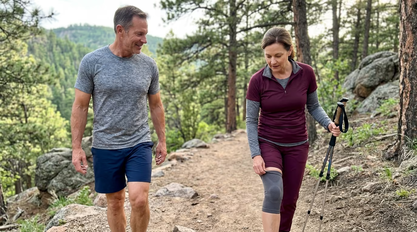

Four structured loading stages build connective tissue capacity, remodel tendon architecture, and protect joint mechanics for lifelong durable movement.





If you have ever searched online for why your knees ache after descending stairs, or why your Achilles tendon feels stiff every morning, you likely found conflicting advice. Some sources tell you to rest completely, while others tell you that your joints are simply wearing out with age.
Neither of these explanations reflects modern connective tissue science. Joint discomfort and tendon sensitivity are rarely problems of simple wear and tear. Instead, they are capacity problems.
When the physical demands of your daily life or training exceed what your tissues can currently handle, irritation follows. Fortunately, tendons, ligaments, and joint structures remain responsive to mechanical load throughout your entire life. By applying the right stimulus in the right dose, you can rebuild tissue capacity, restore movement confidence, and protect your joints for decades of active adventure.
This guide provides a comprehensive framework for joint and tendon health. You will learn how connective tissue adapts to exercise, how to use progressive loading to strengthen vulnerable areas, and how to tell the difference between productive training discomfort and signals that require medical evaluation.
For decades, popular culture treated human joints like the mechanical bearings of an automobile. Under this outdated model, every step on the pavement, every heavy squat, and every mountain descent was thought to grind away finite cartilage until bone met bone.
Modern biomechanics and orthopedics have proven the opposite. Living tissue is dynamic, responsive, and self-renewing. Cartilage, tendons, and ligaments do not degrade purely from use. They remodel and strengthen when exposed to appropriate, progressively managed mechanical stress.
Durable movement is built on the balance between capacity and demand. Capacity refers to the maximum amount of force, speed, compression, and repetitive load a tissue can comfortably handle. Demand is the total stress placed on that tissue by your sport, daily movement, and recreation.
When demand exceeds capacity, tissues become irritated, sensitive, or inflamed. The conventional response is to stop moving entirely until the discomfort disappears. While rest may temporarily reduce symptoms, it also lowers tissue capacity even further. When you eventually return to your favorite sport, your tolerance is lower than when you stopped, creating a frustrating cycle of recurring aches.
The true path to structural durability requires you to systematically expand your tissue capacity. This is achieved not through passive rest, but through controlled, progressive loading. By introducing mechanical tension at manageable volumes, you stimulate cellular remodeling, increase tendon stiffness, and reinforce the muscular structures that protect your joints.
You can learn more about developing lasting physical resilience in our dedicated strength and performance resources.
To train your joints effectively, you must understand how different connective tissues function and adapt. While muscles receive an abundant blood supply and adapt quickly, tendons, ligaments, and cartilage operate on a different biological timeline.
A tendon connects muscle to bone. Its primary role is to transmit high muscular forces to the skeleton and, in many cases, store and release elastic energy like a spring.
A ligament connects bone to bone. Its primary function is to provide passive joint stability and guide healthy movement mechanics.
Articular cartilage coats the ends of bones inside a joint, providing a low-friction gliding surface that distributes compressive forces during movement. Surrounding the entire joint is the joint capsule, lined with synovial membrane that produces lubricating fluid to nourish the joint environment.
Each of these tissues contains specialized cells suspended within an extracellular matrix composed mostly of collagen fibers, water, and proteoglycans. Because these tissues have a lower metabolic rate and blood supply than skeletal muscle, their cellular turnover and remodeling occur over months rather than days.
When discussing tendons, the term stiffness does not mean tightness or a lack of flexibility. In biomechanics, tendon stiffness describes how much force is required to produce a specific amount of tissue deformation.
A stiffer tendon is functionally superior. It transfers force from muscle to bone more rapidly and handles high-velocity energy storage with greater efficiency.
A comprehensive systematic review and meta-analysis published in Sports Medicine examined healthy lower-limb tendon adaptations to exercise. The researchers found that training-induced increases in tendon stiffness are driven primarily by changes in the material properties of the tissue, known as the elastic modulus, rather than large increases in cross-sectional thickness.
This means that a tendon becomes stronger and more resilient primarily by improving its internal collagen architecture and density, not necessarily by getting visibly larger.
When you start a new exercise routine or increase your lifting weights, your muscles adapt rapidly. Within two to four weeks, neuromuscular coordination improves and muscle fibers begin to hypertrophy.
Tendons and ligaments adapt much more slowly. A systematic review published in the British Journal of Sports Medicine demonstrated that significant tendon adaptations require sustained mechanical loading, with interventions lasting longer than 12 weeks showing the greatest benefits.
This discrepancy creates a common vulnerability window. At week six of a new program, your muscles feel strong enough to handle heavier loads or faster running intervals, but your tendons have not yet remodeled their collagen matrix.
If you escalate your training volume based solely on how your muscles feel, you risk overloading your connective tissue. Durable progress requires patience and consistent loading across several months.
Connective tissue responds to three distinct mechanical factors:
First, mechanical stress represents the internal force distributed over the cross-sectional area of the tissue. Heavy resistance training creates high mechanical stress, signaling cells called tenocytes to synthesize new collagen.
Second, strain refers to the percentage of deformation or elongation the tissue experiences relative to its resting length.
Third, strain rate describes how quickly that deformation happens. A slow, controlled heel raise exposes the Achilles tendon to high stress with a very low strain rate. A maximal sprint exposes that same tendon to high stress with an exceptionally rapid strain rate.
High strain rates demand significant elastic capacity. If your training consists only of slow gym exercises, your tendons may lack the reactive capacity needed for sudden accelerations, deceleration, or uneven outdoor terrain.
Every physical activity imposes a specific demand profile on your body. Walking downhill places high compressive and shear forces on the patellofemoral joint. Sprinting for a tennis ball demands rapid force storage and release from the Achilles tendon. Lifting heavy luggage overhead requires stability from the rotator cuff tendons and the glenohumeral joint capsule.
Injury and persistent irritation occur when the immediate demand exceeds current tissue capacity. This concept is illustrated by the capacity-demand gap.
Consider an active individual who walks 8,000 steps per day on flat city sidewalks. Their lower-body tendons have adapted precisely to that level of low-rate, predictable loading.
If that individual travels to the Swiss Alps and completes an intense six-hour mountain hike with 1,500 meters of steep descent, the mechanical demand spikes dramatically. The quadriceps tendon and patellar tendon must repeatedly absorb heavy eccentric deceleration forces on uneven ground.
Because the spike in demand was sudden, the cumulative load surpasses the current capacity of the knee extensors. The following morning, the front of the knee feels hot, stiff, and painful to bend.
This flare-up does not mean the knee joint is permanently damaged. It simply indicates that the tissue was exposed to an unaccustomed workload without adequate preparatory conditioning.
The traditional response to this acute stiffness is complete immobilization and rest. The hiker sits on the couch for two weeks, waiting for all discomfort to vanish.
While rest calms the irritated nerve endings, it also causes cellular deconditioning. Tendon stiffness declines, muscular strength diminishes, and joint cartilage receives less synovial fluid circulation.
When the individual attempts to hike again, their capacity baseline is even lower than before. A smaller physical challenge now triggers the same uncomfortable symptoms.
A far more effective strategy is relative rest combined with active load management. A landmark study on Achilles tendinopathy published in the American Journal of Sports Medicine demonstrated that allowing continued, pain-monitored loading activities did not negatively affect clinical outcomes compared to complete cessation of activity.
By keeping the tissue loaded within tolerable limits, you maintain baseline strength while facilitating structural remodeling. You can review broader strategies for sustaining vitality in our guide to healthy aging and living well.
Building connective tissue durability requires a structured, step-by-step loading progression. You cannot jump directly from complete rest to high-impact jumping without creating irritation.
A proven framework organizes rehabilitation and preparation into four distinct stages. This continuum allows you to match your exercise selection to your current stage of tissue tolerance.
Isometric exercises involve generating muscular force against an immovable resistance without changing the joint angle or lengthening the muscle-tendon unit.
Isometrics are an ideal entry point when a tendon or joint is highly sensitive. They allow you to apply high mechanical tension to the muscle and tendon without the compressive or shear stresses caused by dynamic movement.
Research published in the British Journal of Sports Medicine indicates that isometric loading can produce a meaningful short-term analgesic effect, reducing localized tendon pain for several hours. This makes isometrics an exceptional tool during competitive seasons or when initiating rehabilitation.
A standard clinical dosage for isometric tendon loading consists of:
Common examples include the Spanish squat or leg extension hold for patellar tendon discomfort, the seated single-leg calf raise hold for the Achilles tendon, and the isometric doorway press for the shoulder rotator cuff.
Once isometric holds are tolerated with minimal symptoms, you should progress to heavy slow resistance training. This stage is the cornerstone of long-term tendon remodeling and joint protection.
Heavy slow resistance incorporates both concentric muscle shortening and eccentric muscle lengthening through a controlled, full range of motion. Each repetition is performed with a strict, slow tempo, typically taking three seconds to lift and three seconds to lower.
The systematic review literature on human tendon adaptation confirms that heavy resistance training is the most reliable intervention for improving tendon stiffness, material modulus, and muscular capacity across all age groups.
A practical heavy slow resistance progression follows these parameters:
For the lower body, foundational exercises include slow barbell squats, leg presses, Romanian deadlifts, and seated or standing calf raises. For the upper body, slow dumbbell bench presses, chest-supported rows, and overhead presses build resilient shoulder and elbow tendons.
Heavy slow resistance builds remarkable tissue strength, but it does not prepare your connective tissue for the rapid stretch-shortening cycles of running, jumping, and cutting.
Stage 3 introduces fast elastic demands. In this phase, the tendon must rapidly catch, store, and release mechanical energy like a dynamic spring.
You should only introduce energy storage exercises after you can perform heavy slow resistance training without next-day pain or stiffness.
Energy storage training progresses systematically:
Perform energy storage sessions two times per week, allowing at least 48 to 72 hours between workouts to allow the collagen matrix to recover.
The final stage bridges the gap between structured gym training and unrestricted participation in your chosen sports, whether that involves tennis, trail running, or alpine skiing.
Return to activity is not a single calendar date. According to the international consensus statement on return to sport published in the British Journal of Sports Medicine, physical readiness exists along a continuum.
In this stage, you reintroduce open-ended, reactive movements:
By moving through these four stages in order, you ensure that every layer of tissue capacity is restored before exposing your body to maximum physical demand.
One of the most difficult challenges for active adults is distinguishing between normal training discomfort and symptoms that indicate a worsening condition. Avoiding all discomfort is unnecessary and often counterproductive. However, ignoring sharp warning signs leads to chronic overuse injuries.
The most reliable tool for navigating this balance is the 24-hour response rule.
Tendon and joint tissues often exhibit delayed sensitivity. An exercise session might feel completely comfortable while you are warmed up, only to produce significant throbbing and stiffness twelve hours later.
Because of this delayed response, you must evaluate how your body feels during the session, immediately following the workout, and especially the next morning.
Discomfort is generally considered acceptable if:
Conversely, symptoms suggest an excessive workload if:
To manage your training without complex formulas, use this simple decision model:
Your symptoms are minimal, movement mechanics are clean, and morning stiffness is negligible. You are tolerating the current workload well. You may maintain your current training volume or make a small, progressive increase in resistance or intensity.
You notice elevated tenderness during the workout, or morning stiffness lasts 20 to 30 minutes the next day. Movement mechanics remain intact, but the tissue is showing early signs of fatigue.
Do not increase the training load. Maintain your current level or reduce volume by 20 to 30 percent until next-day symptoms settle back to green.
You experience sharp, stabbing pain, sudden functional loss, joint instability, or severe next-day inflammation. Stop the provocative activity immediately. Substitute gentle, non-irritating movement and seek an assessment from a qualified physical therapist or sports physician.
Using this response model allows you to stay active while keeping tissue stress within safe biological boundaries.
Osteoarthritis is widely misunderstood. Many adults believe that a diagnosis of joint wear means their athletic days are over. In reality, modern medical guidelines view therapeutic exercise as the primary non-pharmacological treatment for maintaining joint longevity.
Clinical guidance from the National Institute for Health and Care Excellence (NICE) recommends structured physical exercise for individuals with osteoarthritis. This guidance specifically includes both local muscular strengthening and general aerobic activity.
Articular cartilage lacks a direct blood supply. It relies entirely on the cyclical compression and decompression of joint movement to draw in synovial fluid, which delivers oxygen and vital nutrients while clearing waste products.
Complete inactivity starves cartilage of this fluid exchange, leading to stiffening and matrix thinning. Gentle, progressive loading acts as a biological pump, nourishing joint surfaces and maintaining range of motion.
NICE clinical reviews emphasize that exercise may initially cause mild, temporary discomfort. However, adhering to a consistent routine produces significant long-term reductions in joint pain and substantial improvements in everyday physical function.
Your muscles serve as the primary active suspension system for your skeleton. When you walk, jump, or land, your muscles contract eccentrically to absorb kinetic energy.
If the muscles surrounding a joint are weak or fatigue rapidly, a greater proportion of that impact force is transferred directly into the passive joint capsule, ligaments, and subchondral bone.
For the knee joint, strong quadriceps, hamstrings, gluteals, and calf muscles act as integrated shock absorbers. Strengthening these surrounding muscle groups reduces localized joint contact stress and optimizes tracking.
A well-rounded joint preservation program includes:
The World Health Organization physical activity guidelines recommend that all adults achieve 150 to 300 minutes of moderate-intensity aerobic exercise weekly, combined with muscle-strengthening activities on two or more days per week. Meeting these baseline targets provides the foundation for durable joint health.
Ligaments provide passive stability, but they rely heavily on neuromuscular coordination to protect them from injury. When you cut or change direction, your nervous system must activate muscles in milliseconds to stabilize the joint.
Neuromuscular training programs integrate progressive strength, plyometrics, balance drills, and movement control mechanics. A meta-analysis published in the American Journal of Sports Medicine demonstrated that comprehensive neuromuscular training programs reduce anterior cruciate ligament (ACL) injury risk by 50 percent overall, and reduce non-contact ACL injuries in female athletes by 67 percent.
Another evidence review in the British Journal of Sports Medicine reported that structured neuromuscular training reduced ACL injury incidence from 1 in 54 down to 1 in 111 among participating athletes.
Building joint longevity is not about making a single ligament unbreakable. It is about training the surrounding muscles and nervous system to control landing forces, manage deceleration, and keep joint angles within safe functional limits.
To protect your joints effectively, you must discard several pervasive myths that lead to poor training decisions and unnecessary anxiety.
Pain is a complex neurological alarm system designed to protect you from potential harm. However, the volume of that alarm does not always match the physical state of your tissues.
You can experience significant tendon sensitivity with very little structural change, or have visible cartilage thinning on an MRI while remaining completely pain-free. Relying entirely on pain as a measurement of structural damage leads to fear of movement. Focus on functional capacity, movement quality, and the 24-hour response rather than chasing immediate zero-pain sensations.
When a tendon feels tight or achy, the instinct is often to stretch it aggressively. However, stretching creates high tensile strain and can compress the tendon against bony prominences, often worsening insertional tendinopathy.
Tendons do not need passive lengthening; they need load tolerance. The systematic review literature consistently demonstrates that resistance training, not passive stretching, is the primary driver of improved tendon stiffness and material remodeling.
The wellness market heavily promotes collagen powders and joint supplements as rapid fixes for aching tissues. However, high-quality clinical research urges caution regarding these claims.
A double-blind randomized controlled trial published in the European Journal of Applied Physiology investigated specific collagen peptide supplementation combined with resistance training in healthy active males. The researchers found that collagen supplementation did not enhance patellar tendon cross-sectional area or mechanical adaptations compared to a placebo.
Nutritional support cannot replace the physical stimulus of mechanical tension. Tendon cells require the direct mechanical stress of resistance exercise to trigger collagen synthesis and structural remodeling.
Many active adults receive an MRI scan showing minor meniscus fraying, disc bulges, or mild tendon degeneration, and assume they must permanently restrict their active pursuits.
Numerous radiological studies show that these structural findings are exceptionally common in completely healthy, pain-free athletes. An imaging scan provides a structural snapshot, but it does not measure functional strength, movement control, or load tolerance. Medical decisions should always prioritize your clinical presentation and movement capacity over an isolated scan report.
The popular guideline that you should never increase training volume by more than 10 percent per week is overly simplistic. For a beginner running three miles per week, adding 10 percent represents an insignificant fraction of a mile. For an experienced marathoner running 70 miles per week, adding 10 percent can easily overload connective tissue.
Progress should be dictated by your individual capacity, training history, and next-day symptom recovery. Adjust one variable at a time, such as distance, speed, or terrain, rather than changing multiple training demands simultaneously.
Active travel places unique, unpredictable demands on your musculoskeletal system. A week of heli-skiing, a multi-day trek across the Scottish Highlands, or navigating cobblestone streets with heavy luggage can challenge unconditioned connective tissue.
Preparing your joints for adventure requires deliberate, task-specific conditioning in the weeks leading up to your departure.
The primary cause of joint distress during mountain trekking is prolonged downhill walking. Descending steep gradients requires continuous, high-volume eccentric contractions from the quadriceps and patellar tendon to brake your body weight against gravity.
To prepare for mountain travel, your training should emphasize eccentric knee extensor strength:
Building this specific eccentric buffer ensures that your knees can handle thousands of downhill steps without developing acute patellofemoral irritation.
Long-haul transit imposes prolonged immobility, cramped joint positions, and circadian disruption, all of which impair soft tissue recovery and muscular readiness.
After a grueling thirty hour transit to Tokyo, I realized my old strategy of just powering through was no longer working. I felt foggy for three days, and my lower back and hips felt locked and irritable. I started digging into circadian biology and realized that timing my light exposure and fasting during the flight could completely shift my recovery.
Now, I never board a long haul flight without a precise schedule for when to eat and when to put on an eye mask. It is the difference between losing a week of your trip and hitting the ground running.
When traveling across time zones:
To deepen your understanding of how recovery protocols optimize physical performance, read through our recovery and sleep strategies and our library of travel and adventure performance guides.
Building durable joints and tendons does not require hours of daily physical therapy. It requires a consistent, sensible commitment to basic progressive loading principles.
You can learn more about our philosophy on lifelong movement capacity by visiting the WealthAtPlay home resource library.
Here is a practical checklist of steps you can take this week to begin reinforcing your connective tissue:
Durable movement is an ongoing process of expanding your physical boundaries through smart, progressive mechanical loading. By treating your connective tissues as living, adaptable structures, you ensure that your body remains capable, resilient, and ready for whatever adventure comes next.
Stay connected for research and practical guidance on longevity, strength, recovery, energy and active living. New ideas to help you stay capable, curious and ready for more.


Explore practical guidance on strength, recovery, energy, travel and longevity for a life that stays active.
explore the Blog